
Bacterial precipitation electron microscopy testing service

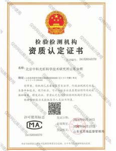

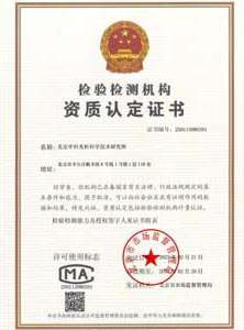

Commitment: Our testing process strictly follows international standards and specifications to ensure the accuracy and reliability of results. Our laboratory facilities are fully equipped with the latest instruments and leading analytical methods. We strictly control every step, from sample collection and processing to data analysis, to ensure clients receive trustworthy test results.


Bacterial Precipitation Electron Microscopy Testing Service – High‑Resolution Structural Characterisation and Morphological Analysis of Bacterial Aggregates, Precipitates and Biofilms
As an ISO/IEC 17025 accredited contract testing laboratory, we offer specialised bacterial precipitation electron microscopy (EM) testing services to pharmaceutical companies, biotechnology firms, clinical microbiology laboratories, environmental monitoring agencies, and research institutions. Bacterial precipitation – the aggregation and sedimentation of bacterial cells, their extracellular polymeric substances (EPS), or antigen‑antibody complexes – is a critical phenomenon in vaccine manufacturing, bioprocess development, wastewater treatment, and diagnostic assay design. High‑resolution imaging is essential to characterise the morphology, distribution, and structural integrity of these precipitates, as well as to detect contaminants, impurities, and morphological abnormalities. Our EM platform integrates scanning electron microscopy (SEM), transmission electron microscopy (TEM), and cryo‑electron microscopy (cryo‑EM) with advanced sample preparation techniques (negative staining, ultrathin sectioning, and freeze‑fracture) to provide a comprehensive structural assessment of bacterial precipitates. All methods are aligned with ISO 13322 (Particle size analysis – Image analysis methods), ISO 21363 (Nanomaterials – Measurement of particle size and shape by transmission electron microscopy), USP <1057> (Scanning Electron Microscopy), EP 2.9.37 (Electron microscopy), and ICH Q6B (Specifications for biotechnological products). Our reports are recognised by the National Medical Products Administration (NMPA), the U.S. Food and Drug Administration (FDA), the European Medicines Agency (EMA), and the World Health Organization (WHO) for regulatory submissions, batch release, and product quality assurance.
Sample Types and Bacterial Precipitate Systems We Analyse
Our electron microscopy laboratory handles a wide variety of bacterial precipitate samples from diverse sources and application contexts. Typical test articles include:
- Vaccine and antigen precipitates – bacterial cell fractions, outer membrane vesicles (OMVs), and protein‑adjuvant precipitates used in subunit and conjugate vaccines
- Fermentation broth precipitates – bacterial cell pellets, inclusion bodies, and co‑precipitated media components from bioprocesses
- Biofilm and extracellular polymeric substance (EPS) precipitates – aggregated bacterial cells, exopolysaccharides, and nucleic acid complexes from environmental and clinical biofilms
- Antigen‑antibody precipitates (immunoprecipitates) – complexes formed during immunoassays, diagnostic tests, and immunoprecipitation protocols
- Bacterial aggregates and flocs – from wastewater treatment, bioremediation, and microbial flocculation processes
- Inclusion bodies and protein aggregates – from recombinant bacterial expression systems
- Contaminated and adventitious agent precipitates – for detection of viral particles, mycoplasma, and other contaminants co‑precipitated with bacterial cultures
- Reference and quality control samples – for method validation, instrument calibration, and proficiency testing
Sample Preparation – Critical Step for High‑Resolution Imaging
We employ a range of sample preparation techniques tailored to the nature of the bacterial precipitate and the imaging mode, ensuring that the native structure is preserved while achieving sufficient electron density and contrast.
- Negative staining (for TEM and SEM) – rapid screening of morphology – For isolated bacterial precipitates (e.g., cell fragments, OMVs, protein aggregates), we use a drop‑casting method on Formvar‑ or carbon‑coated grids, followed by staining with uranyl acetate, phosphotungstic acid, or ammonium molybdate. This technique provides high‑contrast, high‑resolution images of surface morphology, particle size, and aggregation patterns.
- Ultrathin sectioning (for TEM) – detailed internal structure of precipitates and biofilms – For larger precipitates (e.g., biofilm sections, bacterial flocs, and embedded aggregates), we perform fixation (with glutaraldehyde and osmium tetroxide), dehydration, resin embedding, and ultramicrotomy (60‑100 nm sections). The sections are stained with uranyl acetate and lead citrate to enhance contrast of internal membranes, EPS, and extracellular structures.
- Cryo‑electron microscopy (cryo‑EM) – near‑native state imaging for labile precipitates – For samples that are sensitive to dehydration, chemical fixation, or staining (e.g., fragile OMVs, antibody‑antigen complexes, or labile protein aggregates), we use vitrification (plunge‑freezing) in liquid ethane and imaging at cryogenic temperatures (‑180 °C) with a cryo‑TEM. This method preserves the native hydration and structure, enabling accurate measurement of particle size and morphology without artefact.
- Freeze‑fracture and freeze‑etch (for TEM) – visualisation of internal membrane structures – For bacterial precipitates with complex membrane systems (e.g., OMVs, membrane‑associated precipitates), we employ freeze‑fracture and freeze‑etch techniques to expose internal membrane surfaces for imaging.
- Critical point drying (for SEM) – preserving 3D architecture for surface imaging – For bacterial precipitates that are to be imaged by SEM, we perform dehydration (through graded ethanol) and critical point drying (with liquid CO₂) to minimise shrinkage and distortion. The dried samples are sputter‑coated with gold‑palladium or platinum to provide conductivity and enhance the secondary electron signal.
Scanning Electron Microscopy (SEM) – Surface Morphology and Topography
- Surface morphology of bacterial precipitates – imaging of overall structure – Using a field‑emission SEM (FE‑SEM) with a resolution down to 1 nm, we capture high‑magnification images (up to 100 000×) of the precipitate surface. We assess: (a) particle shape and texture – spherical, rod‑shaped, irregular, or aggregated; (b) surface features – the presence of fibrils, vesicles, or biofilm‑like structures; (c) size distribution – the range of particle sizes within the precipitate, including sub‑micron aggregates and large flocs; and (d) porosity – the presence of voids, channels, or porous structures that may affect sedimentation or filtration performance.
- Elemental analysis – Energy‑Dispersive X‑ray Spectroscopy (EDS) for compositional mapping – ASTM E1508 / ISO 22309 – Using EDS detectors integrated with the SEM, we perform semi‑quantitative elemental analysis of the precipitate to identify: (a) mineral precipitates (e.g., calcium phosphate, iron oxides) co‑precipitated with bacterial cells; (b) adsorbed metals – the presence and distribution of metal ions (e.g., Cu, Zn, Pb) bound to the precipitate; (c) chemical gradients – the distribution of elements within a floc or aggregate. EDS mapping provides a spatial map of elemental composition, helping to distinguish between biological and inorganic components.
- Backscattered electron (BSE) imaging – for compositional contrast – BSE imaging provides contrast based on atomic number (Z‑contrast), enabling the differentiation of biological (low Z) and mineral (high Z) precipitates. This is useful for detecting inorganic contaminants or precipitates formed during biomineralisation processes.
Transmission Electron Microscopy (TEM) – Internal Structure and Ultrastructural Details
- Negative staining TEM – rapid screening of particle morphology and size – ISO 21363 / USP <1057> – We examine negatively stained bacterial precipitates at magnifications of 10 000‑100 000× to assess: (a) the morphology of individual particles (e.g., spherical, rod‑shaped, cup‑shaped) and any structural features (e.g., membranes, pili, flagella); (b) particle size distribution – measuring the diameter or length of at least 100 particles per field to generate a size histogram; (c) aggregation state – the degree of clumping or dispersion; and (d) the presence of contaminants – viral particles, sub‑micron debris, or amorphous aggregates.
- Ultrathin sectioning TEM – detailed internal architecture of precipitates – For larger precipitates (e.g., biofilm sections, bacterial flocs), we image ultrathin sections (60‑100 nm) to visualise: (a) bacterial cell integrity – the presence of intact cells, cell wall thickness, and internal structure (cytoplasm, nucleoid, mesosomes); (b) EPS distribution – the localisation and organisation of extracellular polymeric substances around the bacterial cells; (c) cellular damage – evidence of lysis, membrane rupture, or cytoplasmic condensation; and (d) biofilm stratification – the layering and organisation of cells within the aggregate.
- Cryo‑TEM – imaging of labile precipitates in their native state – For samples that are sensitive to chemical fixation or staining (e.g., OMVs, protein assemblies), we perform cryo‑TEM at temperatures of ‑180 °C, using a low‑dose electron beam to minimise radiation damage. The images are analysed for: (a) membrane bilayer structure – the presence and integrity of lipid bilayers in OMVs or membrane fragments; (b) particle size and shape – accurate measurement of particle dimensions without shrinkage artefact; and (c) the internal structure of protein‑based precipitates – the organisation of subunits in protein aggregates or antigen‑antibody complexes.
Quantitative Image Analysis – Particle Size, Shape and Distribution
- Particle size and shape measurement – ISO 13322 / ISO 21363 – Using dedicated image analysis software (e.g., ImageJ, Fiji, or commercial software packages), we measure the Feret diameter, circularity, aspect ratio, and convexity of individual particles from EM images. The mean particle size and the size distribution are calculated (including D10, D50, D90 percentiles). A Gaussian or log‑normal distribution is fitted to the particle size data to assess uniformity.
- Aggregation and clustering analysis – assessment of dispersion – For samples that are intended to be dispersed (e.g., bacterial precipitates used as vaccines), we assess the degree of aggregation by: (a) counting the number of individual particles versus clusters, (b) measuring the cluster size distribution, and (c) calculating the dispersion index (DI). A high DI (> 0.7) indicates good dispersion, while a low DI (< 0.3) indicates excessive aggregation.
- Porosity and surface area estimation – for flocculated and aggregated precipitates – Using image analysis, we measure the area fraction of voids within the precipitate image and estimate the porosity percentage. This parameter is useful for predicting sedimentation and filtration efficiency.
- Statistical analysis of morphological features – We provide statistical summaries (mean, median, standard deviation, coefficient of variation) for the morphological parameters, and we use statistical tests (e.g., Student’s t‑test, ANOVA) to compare the particle populations in different samples or treatment groups.
Quality Control and Contaminant Detection – Ensuring Purity and Product Integrity
- Detection of viral contaminants in bacterial precipitates – for vaccine and bioproduct safety – Using TEM, we examine negatively stained preparations for the presence of virus‑like particles (VLPs) with diameters of 20‑300 nm. We report the number of virus‑like particles observed per grid square or per unit area, and we provide a classification (e.g., bacteriophage, enveloped virus, non‑enveloped virus) based on morphology and size. A finding of > 1 virus particle per 10 grid squares (or an equivalent threshold) is reported as a contamination event.
- Detection of mycoplasma and other cellular contaminants – TEM is used to screen for mycoplasma – which appear as small (150‑300 nm) pleomorphic cells – and for other cellular debris, such as membrane fragments, vesicles, and cellular debris, that may co‑precipitate with the target material.
- Morphological integrity of bacterial cells – assessing the effect of processing and storage – For bacterial precipitates used as vaccines or live biotherapeutics, we assess the morphological integrity of the bacterial cells, including the presence of intact flagella, pili, and a smooth cell surface. Cell shrinkage, lysis, or surface blebbing is scored as a quality defect.
- Detection of amorphous precipitates and non‑biological contaminants – We identify and report the presence of non‑bacterial, non‑biological precipitates (e.g., salt crystals, polymer fragments, or glass particles) that may have been introduced during the manufacturing or formulation process.
Specialised Applications – Bioprocess Monitoring, Vaccine Development and Environmental Analysis
- Monitoring bacterial precipitation during fermentation – for bioprocess optimisation – We analyse samples taken at different time points during fermentation to track the formation of precipitates and to correlate morphological changes with process parameters (e.g., pH, temperature, nutrient depletion). This helps in optimising the harvest time and the product recovery process.
- Characterisation of outer membrane vesicles (OMVs) and bacterial ghost preparations – for vaccine development – We characterise the size, morphology, and integrity of OMVs and bacterial ghosts (empty cell envelopes) by TEM, providing data on their homogeneity and structural stability.
- Biofilm precipitation analysis – for environmental microbiology and wastewater treatment – We analyse biofilm precipitates from wastewater treatment plants, bioreactors, and natural environments, assessing the structure, porosity, and the presence of filamentous bacteria and mineral precipitates.
- Quality control of bacterial biopesticides and biofertilisers – We assess the morphology, size, and purity of bacterial precipitates used as biopesticides or biofertilisers, verifying that they meet product specifications and are free from contaminants.
Data Interpretation and Reporting – Comprehensive Documentation
Our final report provides a complete, transparent and actionable record of the electron microscopy analysis. The report includes:
- Sample information – unique sample ID, source, preparation history, and the purpose of the analysis
- Methods summary – a description of the EM mode (SEM, TEM, cryo‑EM), the sample preparation technique, the imaging parameters (accelerating voltage, working distance, spot size), and the analytical software used
- Representative images – high‑resolution images (JPEG or TIFF format) at multiple magnifications, with scale bars and explanatory annotations
- Quantitative data – particle size distribution (histograms), shape parameters (aspect ratio, circularity), aggregation index, porosity percentage, and contaminant counts
- Statistical summary – mean, standard deviation, coefficient of variation, 95 % confidence intervals, and statistical comparisons (where applicable)
- Interpretation and conclusions – a clear statement on the morphology, uniformity, purity, and structural integrity of the bacterial precipitate, with a discussion of the relevance of the findings to the client’s application (e.g., bioprocess optimisation, vaccine quality, or contaminant detection)
- Raw data and supplementary materials – all raw images (including fields of view that are not included in the main report) and the primary data files (e.g., particle size measurement files) are provided in digital format
Quality Control, Instrument Calibration and Proficiency Testing
To ensure the accuracy, reproducibility and regulatory compliance of our EM data, we follow strict quality control and instrument calibration procedures:
- Instrument calibration and performance verification – SEM, TEM, and cryo‑TEM are calibrated on a regular basis using certified reference materials (e.g., gold or carbon grids with known lattice spacings) to ensure that the magnification, resolution, and image distortion meet the manufacturer’s specifications.
- Magnification accuracy – using grating replica or calibration grids – We use calibration grids (e.g., with 2 160 lines/mm or 28 800 lines/mm) to verify the magnification accuracy; the error is maintained at ≤ 2 %.
- Energy dispersive X‑ray (EDS) calibration – with certified standards – The EDS system is calibrated using certified reference materials (e.g., pure elements or known alloys) to ensure accurate elemental identification and quantification.
- External proficiency testing – we participate in EM proficiency testing schemes (organised by NIST, UK NEQAS, or other providers) to validate the accuracy of our morphological analysis and to benchmark our performance against other laboratories.
- Image archiving and data integrity – all raw images and analytical data are stored in a secure, version‑controlled database with a full audit trail (including the date, time, operator, and instrument settings) to support regulatory compliance.
Report Acceptance and Regulatory Compliance
All bacterial precipitation EM analyses are performed under our ISO/IEC 17025 accreditation and in compliance with Good Laboratory Practice (GLP) principles, USP <1057>, EP 2.9.37, and ICH Q6B guidelines. Our reports are accepted by the National Medical Products Administration (NMPA), the U.S. Food and Drug Administration (FDA), the European Medicines Agency (EMA), and the World Health Organization (WHO) for product registration, batch release, and quality assurance. Bilingual (Chinese/English) versions are available to facilitate submissions to national and international regulatory authorities.
Note: Due to business adjustments, we do not accept individual client testing requests.
The above is an introduction about Bacterial precipitation electron microscopy testing service. For further questions, please consult our online engineer.
Laboratory Instruments


Partner Clients

Related Projects
 Hydrogen sulfide corrosion testing service
 Pressure relief valve hydraulic burst testing service
 Freezing experiment
 Ultimate impact torque testing service
 Corrosion resistance testing service
 20℃ impact energy testing service
 Pressure leak testing service
 AC dielectric testing service
 Vibration testing service
 Housing pressure testing service